Anatomy of the Eye
Anatomy of the Eye
The eye is a remarkable organ that allows us to experience the world around us. It functions like a camera, capturing light and transmitting it to the brain, where it is interpreted as images. Understanding the anatomy of the eye is essential for better comprehension of how we see and why eye health is so important.
At Columbia Eye Associates in Lake City, FL, we are dedicated to offering the best possible care for your eyes, from routine exams to advanced treatments. By understanding the intricate parts of the eye, we can help you maintain and improve your vision.
Why is Understanding Eye Anatomy Important?
In Lake City, FL, many individuals seek professional help with their vision, whether it’s for multifocal contact lenses, cataract surgery, or general eye exams. As demand for eye care services increases, it becomes vital for patients to understand the structure of the eye and how it affects vision.
With knowledge of the eye’s anatomy, patients can better understand how different conditions can affect their sight and how we, at Columbia Eye Associates, can help treat those conditions effectively.
Our goal is to make sure you have a clear understanding of your eye health and how we can support you in maintaining healthy vision.
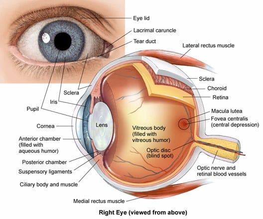
Key Parts of the Eye and Their Functions
The anatomy of the eye includes several important structures, each playing a vital role in how we see. Here are some key components:
Anterior Chamber
The anterior chamber is the front section of the eye, located between the cornea and the iris. It is filled with aqueous humor, a clear fluid that helps nourish the eye and maintain eye pressure. This chamber plays a crucial role in eye health, providing nutrients to the eye’s internal structures.
Aqueous Humor
The aqueous humor is a clear, watery fluid that fills the anterior chamber of the eye. It helps maintain intraocular pressure and delivers essential nutrients to the eye’s tissues. It is constantly produced and drained to ensure that eye pressure remains stable.
Blood Vessels
Blood vessels, including arteries and veins, transport oxygen and nutrients to the eye and remove waste products. Proper circulation is necessary for eye health, as the retina and other internal eye structures depend on a steady supply of blood.
Caruncle
The caruncle is a small, red portion of the corner of your eye. It contains modified sebaceous and sweat glands. Though often unnoticed, this part of the eye helps with lubrication and the removal of debris.
Choroid
The choroid is a layer of blood vessels located between the retina and the sclera. It is responsible for supplying oxygen and nutrients to the retina, keeping the eye’s light-sensitive tissue healthy.
Ciliary Body
The ciliary body produces the aqueous humor and controls the shape of the lens to help focus images. It also contains the ciliary muscles, which help change the shape of the lens to accommodate for near and far vision.
Cornea
The cornea is the clear, dome-shaped surface that covers the front of the eye. It acts as the eye’s protective barrier and plays a key role in focusing light onto the retina, helping you see clearly.
Iris
The iris is the colored part of the eye that surrounds the pupil. It controls the amount of light that enters the eye by adjusting the size of the pupil. This structure plays a role in regulating vision and protecting the retina from excessive light.
Lens (Crystalline Lens)
The lens is a transparent, flexible structure located behind the iris. It helps focus light onto the retina by changing its shape. This process, known as accommodation, allows us to focus on objects both near and far.
Lower Eyelid
The lower eyelid is the skin that covers the bottom of the eyeball when closed. It helps protect the eye from debris and provides moisture through the tear film.
Macula
The macula is the central part of the retina responsible for sharp, detailed central vision. It enables us to read, recognize faces, and see fine details clearly. Damage to the macula can lead to conditions like macular degeneration, which affects vision.
Optic Nerve
The optic nerve is a bundle of nerve fibers that transmits visual information from the retina to the brain. It plays a vital role in the interpretation of light, dark, and color, allowing us to see.
Posterior Chamber
The posterior chamber is located behind the iris and in front of the lens. It also contains aqueous humor, which helps maintain the shape of the eye and facilitates nutrient delivery to the internal structures.
Pupil
The pupil is the circular opening in the center of the iris that allows light to enter the eye. The size of the pupil adjusts based on light conditions to help regulate how much light enters the eye.
Retina
The retina is the light-sensitive layer at the back of the eye. It contains photoreceptor cells that detect light and convert it into electrical signals, which are then sent to the brain via the optic nerve. The retina is essential for clear vision.
Sclera
The sclera is the white, outer layer of the eye. It helps maintain the shape of the eye and protects its internal structures. The muscles that control eye movement are attached to the sclera.
Suspensory Ligament of Lens
This series of fibers connects the ciliary body to the lens, holding the lens in place. It helps the lens change shape during accommodation, which allows the eye to focus on objects at various distances.
Upper Eyelid
The upper eyelid is the movable skin fold that covers the front of the eye when closed, including the cornea. It helps protect the eye and facilitates the spread of tears across the surface of the eye.
Vitreous Body
The vitreous body is a clear, jelly-like substance that fills the back of the eye, between the lens and retina. It helps maintain the shape of the eye and supports the retina in its position.
If you have any questions about the anatomy of the eye or need assistance with your eye health, don’t hesitate to contact us today.